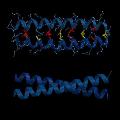
Figure 1.10. Lateral views of the GCN4 leucine zipper dimer (based on Fig. 3B & C of O'Shea et al. (1991); PDB deposition code ZTA2, Bernstein et al. (1977)).
(A) The main chains are displayed as ribbons and the sidechains of the residues involved in the 4-3 hydrophobic repeat, within the dimer interface, are represented by balls and sticks. This illustrates how these residues pack side-by-side against one another, as opposed to the interdigitation model of Landschulz et al. (1988). From this perpective, the molecule resembles a ladder, in which the sides are formed by the helical backbones and the rungs are formed by the hydrophobic sidechains. (B) View perpendicular to (A) which shows how the main chains (displayed as ribbons) cross one another at an angle of about 18 . Figure prepared with the program MOLSCRIPT (Kraulis, 1991).