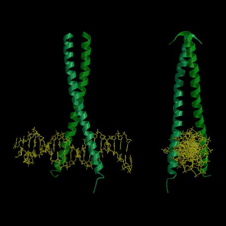
Figure 1.12. X-ray crystal structure of the GCN4-bZIP:AP-1 complex (based on Fig. 3 of Ellenberger et al. (1992); PDB deposition code YSA1, Bernstein et al. (1977)).
(A) View perpendicular to both the DNA and coiled-coil axes. The GCN4-bZIP monomers form continuous smooth  -helices which combine at their C-termini to form a coiled-coil and then gently diverge to allow the basic regions to lie in the major groove of the AP-1 site, resulting in a Y-shaped molecule. The DNA within the complex is straight and in the B form. (B) The view down the DNA axis shows how the GCN4-bZIP dimer grasps the DNA like a pair of -helical tweezers. Figure prepared with the program MOLSCRIPT (Kraulis, 1991).