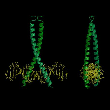
Figure 1.13. X-ray crystal structure of the GCN4-bZIP:ATF/CREB complex (based on Fig. 2 of König and Richmond (1993); PDB deposition code DGC1, Bernstein et al. (1977)).
(A) View normal to both the DNA and coiled-coil axes. The structure is very similar to the GCN4-bZIP:AP-1 complex with the exceptions that the protein monomers are crystallographically symmetrical and that the DNA within the ATF-CREB site is kinked to accommodate binding of the basic domains. (B) Orthogonal view to (A) which shows that GCN4-bZIP straddles the ATF/CREB site in a similar fashion to the way it does when bound to the AP-1 site. Figure prepared with the program MOLSCRIPT (Kraulis, 1991).