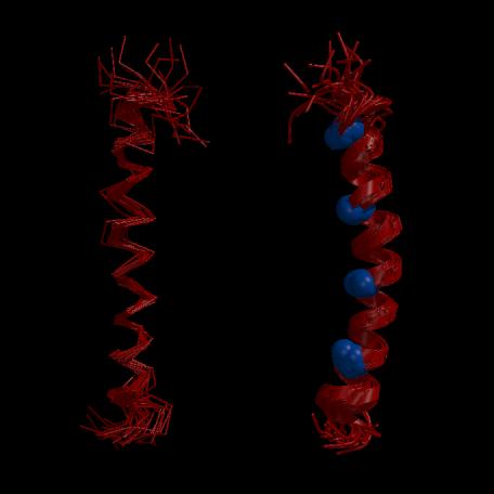
Figure 1.7. NMR solution structures of the GCN4 leucine zipper (Saudek et al., 1991b; PDB deposition code ZTA1, Bernstein et al., 1977).
(A) Lateral view of a C trace of 20 structures of the GCN4 leucine zipper superimposed for minimum RMSD between backbone N, C
 and C atoms. These structures show that the leucine zipper of GCN4 adopts a well defined extended
  -helix which diverges slightly at both termini. (B) View orthogonal to (A) with the molecule represented as an
  -helical ribbon and the C
 atoms of the interfacial leucine residues drawn as spheres. The helix forms a gentle curve along its length, with the leucine residues aligning along the concave side. Figure prepared with the program MOLSCRIPT (Kraulis, 1991).

