3. Give the structural informations of this PDB file (a) number of protein chains, (b) number of helix in one protein chain, (c) the first and last residues in the helix 6 .
Protein Structure Database -PDB-.
1. Search human Mlh1 or its homologous proteins in Protein Data Bank (PDB) database. You just need to search for protein structure, do not find complex structure. Give its (a) ID number, (b) Name of the molecule, (c) experimental method for structure determination and (d) Resolution of the structure.
Ans: ID number: 1BKN
Name of the molecule: Crystal Structure Of An N-Terminal 40Kd Fragment Of E. Coli DNA Mismatch Repair Protein Mutl
Experimental method for structure determination: X-ray Diffraction
Resolution of the structure: 2.90
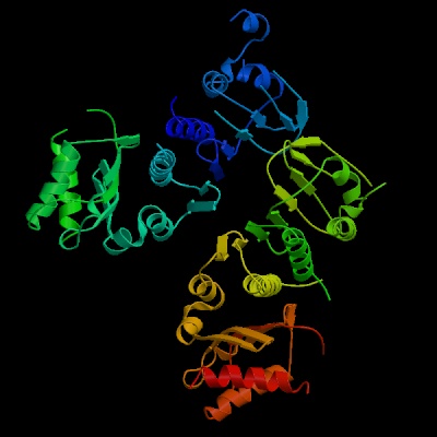
2. Show its structure in ribbons form (400x400).
Ans:
3. Give the structural informations of this PDB
file (a) number of protein chains, (b) number of helix in one protein chain, (c)
the first and last residues in the helix 6 .
Ans: Number of protein chains: 2
Number of helix in one protein chain:
Chain 1BKN:A - Number of Alpha 9
Chain 1BKN:B - Number of Alpha 8
The first and last residues in the helix 6:
Chain 1BKN:A first residue : K last residue: C
Chain 1BKN:B first residue :T last residue:Q
4. Give the Class, Fold, Superfamily and Family of this protein in Structural Classification of Proteins (SCOP).
Ans: Class: Mainly antiparallel beta sheets (segregated alpha and beta regions)
Fold: ATPase domain of HSP90 chaperone/DNA topoisomerase II/histidine kinase8-stranded mixed beta-sheet; 2 layers: alpha/beta
Superfamily: ATPase domain of HSP90 chaperone/DNA topoisomerase II/histidine kinase
Family: DNA gyrase/MutL, N-terminal domain
5. (a) Show Ramachandran plot for this PDB file. (b) How many residues are in the most favoured regions of Ramachandran plot?
Ans:
166 residues are in the most favoured regions of Ramachandran plot.