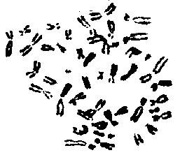
 During
this chromosome lab, the students prepare,
develop and stain their won chromosomes. The metaphase
spread is photographed and the students then do the
karyotyping of their own chromosomes. Students will get a
feeling of accomplishment and excitement over the
numerous, but simple steps that
are required.
A note of caution: this lab requires the withdrawal of blood from the arm by a qualified
individual. The overview of the lab procedure is rather straightforward. A blood sample is taken
and white blood cells grown in a special medium for three days under the influence of the mitotic
stimulant, phytohemaglutinin. After 72 hours, Colcemid, a chemical that inhibits spindle
formation, is added. The red blood cells are exploded with a hypotonic solution of potassium
chloride. The remaining swollen white blood cells are fixed with alcohol and acetic acid. The
resulting cells are dropped from a pasteur pipette onto a clean slide, causing the cells to splatter
and separate. When the slide is stained with giemsa stain, the chromosomes of the individual
white blood cells are visible (Macgregor & Narley 1983).